


For Brain Trauma, an Emerging Suite of Diagnostic Gadgets
The voice of the 911 dispatcher crackled over the intercom at the Colorado Springs Fire Department. A 73-year-old man at a memory care unit in a nursing home had fallen and might have sustained a head injury. In short order, Dr. Stein Bronsky, the medical director, sped to the scene aboard a fire engine.
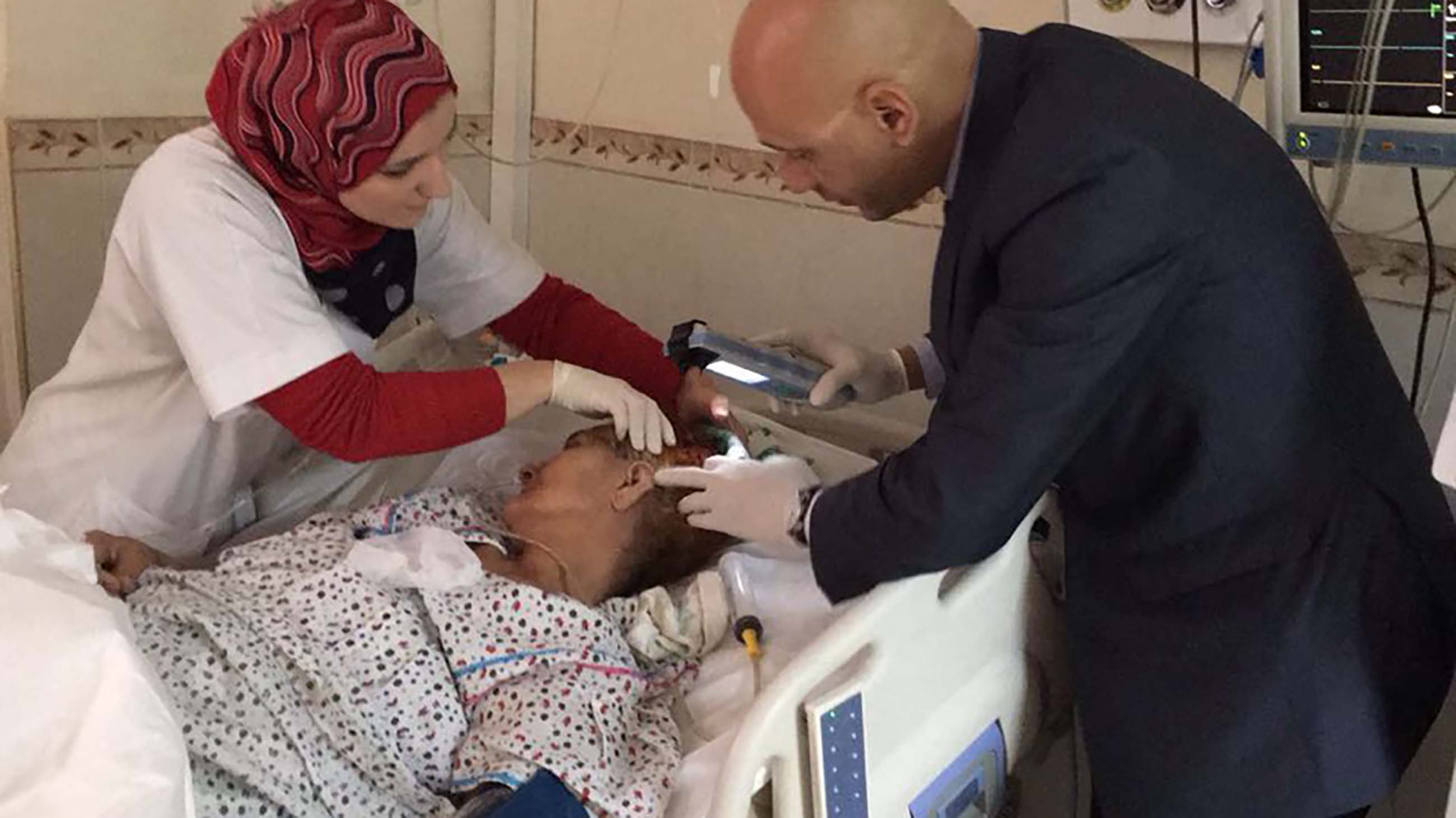
Upon arrival, he and an ambulance crew found the man sitting upright in a chair in a common area. They quickly learned that no one had witnessed the accident and that he was taking blood thinners. As the paramedics started to assess vital signs and check for any external bleeding, Bronsky produced the Infrascanner, a portable device that uses near-infrared light to detect bleeding inside the brain. About two minutes later — after taking eight measurements of the right and left sides of the frontal, temporal, parietal, and occipital areas of the man’s head — the result was positive.
As a precaution, Bronsky repeated the test with the same results.
“Based on that positive reading, knowing that certain hospitals have neurosurgery capability and some don’t, I instructed the crew to bring the patient not to the closest hospital but to the closest one that had the neurosurgeon,” he recalls.
That decision made all the difference, and Bronsky credits that portable scanner, which allowed him to make an informed and speedy decision at the scene. Without the device, Bronsky says the nursing home resident would likely have been transported to the closest hospital for a CT scan, and when the result came back positive, he would have had to be moved to yet another hospital with neurosurgical capability — a process that’s more time consuming and expensive, and when dealing with head injuries, when time to treatment can be crucial, more risky for the patient.
This year, the Colorado Springs Fire Department is preparing to embark on a study using three of the devices to determine whether paramedics will integrate the device fully into their triage routine.
The Infrascanner, manufactured by Philadelphia upstart InfraScan, Inc., gained approval from the Food and Drug Administration in 2011 and it is one of a number of new technologies that have been developed, or are in the pipeline, to assess a variety of head traumas — many of them time-sensitive — from concussion and traumatic brain injury, to strokes. The devices are aimed at medical technicians and doctors toiling on the battlefield, on the sports field, in ambulances and operating rooms, as well as in remote locations where there is no easy access to large and expensive imaging equipment like CT and MRI scanners.
High-tech companies are racing to develop an array of handheld gadgets, tests, and other novel solutions. BrainScope’s Ahead 300 and ElMindA’s BNA harness the power of electroencephalograms (EEGs). Third Eye Diagnostics’ Cerepress relies on eye pressure. Banyan Biomarkers and Grace Laboratories are creating biomarker blood tests. Neuropsychology tests developed by the military are also part of the mix in addition to ImPACT Applications’ immediate post-concussion assessment and cognitive testing and AnthroTronix’s DANA software’s computerized cognitive tests.
This emerging ecosystem of portable technologies has the potential to enhance our understanding about how to treat and mitigate the effects of head trauma. Handheld technology gives medical personnel a critical tool for early diagnosis of TBI in the field. It can expedite surgery, when required, save more lives, and prevent neurological damage. Despite great advances in medical research, no handheld device is capable of accurately diagnosing every variety of head injury. Nevertheless, the market for such products is huge both in the U.S. and overseas — especially in rural and remote communities where advanced imaging technology remains beyond their financial and logistical reach.
“If you have a structural lesion in your brain or blood in your brain, you are sort of out of the concussion domain and into the traumatic brain injury domain,” says Dr. Matthew Kirschen, an attending physician in critical care medicine at the Children’s Hospital of Philadelphia. With moderate to severe TBI, things can get complicated quickly. “The time frame over which you need to intervene varies,” Kirschen says, “but the time frame to start the monitoring is as soon as possible.”
According to the BBC, an early version of the Infrascanner was tested in India a decade ago and the company now says hundreds of the devices are in use overseas. Last month, the British and Irish Boxing Authority announced that the Infrascanner will be ringside for their professional boxing events and military forces in Germany, Israel, the Philippines, Spain, and Turkey have deployed it or are evaluating it for future use.
In 2015, the London Air Ambulance charity began trials to screen patients with the device before they arrive at a hospital. According to Infrascan, it’s also being used or tested in India, Indonesia, Poland, Romania, and Russia.
The skull is unique from other body parts in that it’s a rigid box that cannot expand. As blood accumulates in the brain there are four elements in the skull: brain tissue, cerebral spinal fluid, normal blood within blood vessels, and “other things that don’t belong” such as a rapidly expanding hematoma or hemorrhage, Kirschen explains. Blood swirling around where it shouldn’t be is a huge irritant that also causes brain swelling. So, as the hemorrhage starts to build up, it pushes other structures out of the way to make room for itself. This compresses brain tissue and the patient starts to get symptoms of dysfunction. Why? Because the brain stem sits at the center-bottom of the brain and it controls critical bodily functions like breathing and our heart rate. So, as this area of the brain gets squished and stops working it leads to the patient’s demise unless they get immediate medical attention.
Every generation, from toddlers and teenagers to young adults and senior citizens, is at risk for concussion and TBI. Everyone from the Centers for Disease Control and Prevention to the military compiles statistics showing just how frequently these injuries occur. The film “Concussion” shined a spotlight on the NFL, but the threat of head injury is just as real for high school and college athletes as it is for professional athletes.
The CDC says TBI contributes to about one third of all injury deaths in the U.S. TBI accounted for about 2.5 million visits to the ER, hospitalizations, or deaths in 2010 at an economic cost of $76.5 billion. The CDC also cautions that TBIs from sports and recreation have become “a major public health problem in the United States.” Between 2001 and 2009, about 65 percent of such injuries occurred in children and teenagers 19 years old or younger. Most of the TBIs were associated with injuries from bicycling, football, or the playground.
In the military, close to 80 percent of all TBI cases are sustained in the U.S.
“The number one most common cause of TBIs is not what you would actually think — it’s non-combat, fall related,” says Dr. Aaron Brodsky, a clinical neuropsychologist who serves as the TBI advisor at the Wounded Warrior Regiment at the Marine Corps Base Quantico. “It’s people falling down stairs, people tripping on the snow, and the ice in the wintertime.”
The lion’s share — more than 290,000 — are mild TBI, which account for more than 82 percent of cases from 2000 through the first half of 2016, according to the Defense and Veterans Brain Injury Center. Nearly 32,000 (9 percent) are moderate, more than 5,000 (1.4 percent) are penetrating cases, and nearly 3,700 (1 percent) are severe cases.
The brain, like all biological tissue, is permeable to electromagnetic radiation. This is the foundation for X-rays, CT scans, and near-infrared (NIR) imaging.
Claudia Robertson, a neurosurgery professor at Baylor College of Medicine, worked with the late Dr. Britton Chance of the University of Pennsylvania to research ways of measuring oxygen saturation in the brain using NIR. The duo examined patients with TBI and found that NIR had mixed results.
“The common denominator in the patients where the NIR did not work was the presence of intracranial blood,” she says. “This gave us the idea that we might be able to identify the presence of blood using NIR.”
In a 2010 study conducted for FDA approval of the Infrascanner, Robertson, who has no financial ties to InfraScan, described how NIR highlights intracranial hematomas: there’s a dramatic difference in the way extravascular blood — blood outside the vessels — absorbs near-infrared light compared to intravascular blood. In the case of an acute hematoma, extravascular blood typically has 10 times the concentration of hemoglobin than in “brain tissue where blood is contained within vessels.”
Her study of 365 patients with TBI found that the Infrascanner had a sensitivity of 88 percent and specificity of 90.7 percent in detecting intracranial hematomas.
“We concluded that the Infrascanner would be useful in screening patients with traumatic brain injury, to identify those patients at high risk for an intracranial hematoma that might need surgery,” she says. “Of course, it cannot replace the need for a CT scan, which is the standard test for assessing a traumatic brain injury.”
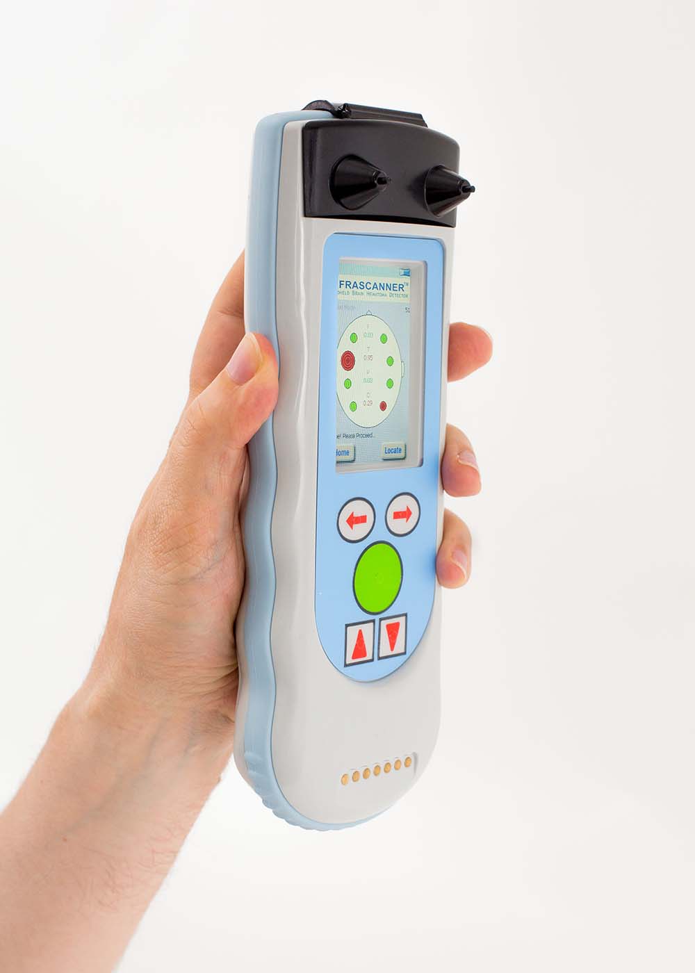
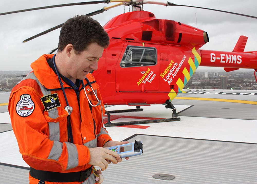
The Infrascanner resembles an oversized smartphone with a small screen and some high-tech features straight out of “Star Trek.”
Visual: InfraScan
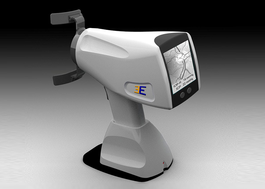
The Infrascanner resembles an oversized smartphone with a small screen and some high-tech features straight out of “Star Trek.” Imagine being a patient in the Enterprise’s sickbay as Dr. McCoy places the scanner at various points on your head to measure hematomas that are greater than 3.5 milliliters in volume and up to 2.5 centimeters deep from the surface of the brain. With the press of a button, it uses a laser to generate the light and there’s a detector with an optical filter on it that captures the near-infrared light that’s scattered through the skull. A disposable fiberoptic shield connects to the device to turn it on. The fiberoptic filaments serve another purpose: They are used like a brush to move hair aside so measurements can be taken directly against the skin of the head without having to shave the patient.
The difference in light absorption between the left and right side of the brain determines if there’s a bleed. The technology requires measuring the same pair — the left and right sides of the frontal, temporal, parietal, and occipital areas of the brain — three times before it confirms a hematoma. Once a scan is completed, the display shows a representation of the skull and a red dot (positive for a hematoma) or a green dot (normal). The size of the dot (small, medium, large) indicates the severity of the bleed.
The Infrascanner was designed with a simple interface so that paramedics can use it in stressful situations.
“It’s like a thermometer. You just measure the temperature,” Baruch Ben Dor, CEO of InfraScan, says. “Here, we just measure if somebody has a bleed.”
The device won’t detect deep brain bleeds. “So, the fact that we’re able to see only an inch into the brain is still enough to detect the vast majority of the bleeds that happen in the case of head trauma,” he says. False positives are possible. But it has a built-in protocol for users to verify a bleed.
Some features, including a dimmable screen and the ability to operate it with disposable batteries, were designed to meet military requirements. The Infrascanner was funded in part by the Office of Naval Research and the U.S. Marine Corps. The device was field tested in Fallujah, Iraq, in 2008. The Marine Corps has since deployed more than 170 of the Infrascanner model 2000 in its standard trauma kit for battalion aid stations.
The Marines have asked InfraScan for the ability to perform non-invasive intracranial pressure monitoring with the device and the integration of a screening tool, a neuropsychology test called the Military Acute Concussion Evaluation (MACE). TBI can cause the brain to swell and this leads to a dangerous rise in pressure. At present, there’s no simple way to measure intracranial pressure in the field, during medical evacuations, or even in the operating room. Ben Dor says InfraScan is developing a new system called the Multifunction Infrascanner Brain Injury Monitor that will include a non-invasive way to measure intracranial pressure. MACE, he says, will be added as future software upgrade to the Infrascanner model 2000.
The Defense Department requires service members to undergo a baseline neuropsychology test before deployment. In addition to MACE, a number of other tests have been developed including ImPACT Applications’ ImPACT, which has FDA approval, the Automated Neuropsychological Assessment Metrics (ANAM), and AnthroTronix’s DANA software.
Dr. Brodsky of the Wounded Warrior Regiment says testing both before and after deployment is critical because many service members do not report injuries.
“It’s hard enough for me as a neuropsychologist to predict where a person should be when I’m doing testing on them. But if I actually have a pre and post-measure then that’s gold,” he says.
Concussions are quite common among children. When it comes to assessing kids with head trauma — whether from a sports injury or a fall — Dr. Joseph Maroon, the team neurosurgeon for the Pittsburgh Steelers and the medical director of the WWE, as well as a clinical neurosurgery professor at the University of Pittsburgh Medical Center, says CAT scans are frequently overused in emergency rooms.
“The concern is whether or not there may be an intracranial hematoma. In the vast majority, this is not so, but to be a thousand percent sure they do a CAT scan,” he explains. “But with the Infrascanner and with a good history and physical, and intelligent parents who can watch the child, CAT scans are not needed as often as they are used.”
Many doctors remain concerned about the radiation exposure from CAT scans.
Dr. Robertson, of Baylor College of Medicine, notes in a recent paper: “NIRS is thought to be more effective in the pediatric population since children have thinner scalps and skulls so there is less noise in the NIRS signal. Moreover, NIRS monitoring may particularly benefit children by allowing them to forgo serial CT imaging for intracranial hemorrhage monitoring, decreasing their overall radiation exposure at a vulnerable age.”
Dr. John Oldershaw, the chief of imaging services at the Pittsburgh Veterans Affairs Healthcare System, says: “A single CT scan of the head is equivalent to 400 oral X-rays or 100 single view chest X-rays.”
This is one of the reasons that the Pittsburgh VA is utilizing the Infrascanner, which emits no radiation, to help triage low-suspicion, elderly patients: “The main utility is patients that have mental status changes or patients that have sustained falls that are either witnessed or unwitnessed,” Oldershaw says.
The hospital — the only one in the VA system to use the Infrascanner — purchased six Infrascanners and two of them are in use at its satellite nursing home facility for veterans.
Oldershaw says one area of concern is among elderly patients with a history of stroke who have encephalomalacia — a condition where the brain may have atrophied in certain areas, resulting in asymmetry. This can set off an alarm bell for a bleed since the Infrascanner looks for symmetry when it takes measurements. Otherwise, he says he’s been very happy with the results: He’s brought the technology to the attention of the leadership of the VA in Washington, D.C., to see if it could be integrated at VA hospitals nationwide.
Injuries suffered by professional football players have helped to drive the market for new diagnostic tools. Here, the BrainScope is featured in a news report on the issue.
Boxing and mixed martial arts are two sports Dr. Maroon says would especially benefit from having a ringside portable device to assess head trauma.
“These are venues where the ultimate goal is to create brain damage either with a concussion or hypoxia so that the incidence of blood clots and brain damage is clearly evident in these particular venues,” he explains.
Maroon says he has used the Infrascanner both at professional sporting events and during office visits to check for intracranial hematomas: “Rather than get a CAT scan, I will do that if the symptoms don’t suggest a more significant problem.”
Overall, he says the Infrascanner’s sensitivity — its ability to “pick up a teaspoon of blood”— provides another tool for doctors to use as part of TBI assessment incorporating a baseline test, medical history, and a physical and neurological exam.
The UCLA BrainSPORT program is part of the NCAA-Department of Defense CARE Consortium —a three-year, $30 million initiative to study sport-related concussions.
Dr. Chris Giza, BrainSPORT’s director and a professor of pediatric neurology and neurosurgery, is researching a variety of concussion detection technologies including helmet and non-helmeted acceleration sensors, brain blood flow monitors, balance sensors, among others. He says UCLA has focused its acceleration research on an earpiece sensor and the helmet-based system called the Head Impact Telemetry System, which measures the force and direction of impact to the helmet to measure force applied to the head and brain.
This year, Giza is gearing up for an NIH-funded study to examine brain blood flow in patients who have had concussions. The study will use Neural Analytics’ portable transcranial Doppler, which is like an ultrasound for blood flow, and compare this with the gold standard — an MRI measure for blood flow called arterial spin labeling, he says.
Two companies have also developed portable technologies for diagnosing head trauma using an EEG, which picks up electrical impulses in the brain by attaching electrodes to the patient’s scalp.
ElMindA, a data-science company based in Israel, has developed an FDA-approved cloud software called BNA, which stands for Brain Network Activation mapping. BNA measures EEG data after a concussion to find changes in electrical function in the brain. “Our goal is to measure neuronal networks, neuronal firing in the brain,” explains CEO Ronen Gadot. BNA is used in concert with a variety of EEG hardware, including Electrical Geodesic’s Sensor Net, which looks like a high-tech hairnet filled with dozens and dozens of electrodes that record brain activity.
BrainScope’s Ahead 300 is a single-use disposable electrode headset that uses EEG measurements to assess mild TBI or concussions. It also has been approved by the FDA and was developed with the Department of Defense, according to a September 2016 press release. Michael Singer, the company’s CEO, did not respond to repeated interview requests from Undark.
While Dr. Giza sees promise in some of the new technologies undergoing rigorous testing, he sounds a cautionary note about technology that gets fast-tracked to consumers. He says there are many “impact sensors that are not going through any validation process or are just going through internal validation at the company, then being marketing directly to parents or schools.”
Our blood has a story to tell. When someone has prostate cancer, or sustains a heart attack or head trauma, biomarkers appear. Decoding the message — and doing so with a portable, hand-held test that can be completed in a matter of minutes — is a now a focal point of TBI biomarker research.
The global market for a mild TBI portable biomarker test is approximately $6 to $9 billion — three times the size of the market for a hand-held stroke test, says Robert Rhinehart, the senior managing member of Grace Laboratories. Grace Laboratories, an acronym for glutamate receptor and common epilepsy, was established to commercialize the research of the lab’s founder and chief neuroscientist, Svetlana Dambinova.
“We are in the process of developing a five-minute sideline test that you can put a drop of whole blood on a strip and in about five minutes you can take a picture of it with your iPhone or put it in a reader and we can determine if someone has sustained a very mild concussion,” Rhinehart says.
Dambinova, who researches neurotoxicity biomarkers and their relevance to cerebral ischemia, epilepsy, substance abuse, and TBI says: “Head impact drives glutamate receptor peptides to be released continuously into the bloodstream through the compromised blood brain barrier within hours to days after impact.”
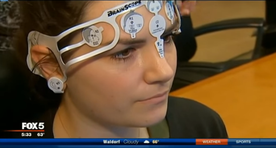
BrainScope’s device is a single-use disposable electrode headset that uses EEG measurements to assess mild brain injuries or concussions. Here the unit is featured in a local news segment.
Visual: YouTube
Grace Laboratories has completed work on a prototype for a handheld test for a stroke with their Russian partner, Skolkovo Biomed and its research company, DRD Biotech.
Meanwhile, Banyan Biomarkers is also racing to develop a blood test.
“Every organ but the brain has a simple blood test,” says Ronald Hayes, co-founder and chief science officer of Banyan. “So it’s a way of assessing organically whether or not there’s injury.”
Banyan’s biomarker research is focused on ubiquitin C-terminal hydrolase L1 (UCHL1), which is found in brain neurons, and glial fibrillary acidic protein (GFAP), which is found in astrocytes, specialized glial cells found in the central nervous system. The company has licensed its biomarker research to a number of medical device makers including Abbott, Philips, and Quanterix to develop a handheld test for TBI.
Unfortunately, there’s no easy way to measure intracranial pressure. A dangerous rise in pressure can occur due to brain swelling from TBI or hydrocephalus. Presently, doctors monitor this is by drilling a hole in the skull and inserting a sensor into the cranium. The procedure isn’t without risks of infection and hemorrhaging.
Third Eye Diagnostics has developed a prototype for a hand-held, non-invasive tool called the Cerepress to measure intracranial pressure by gauging the pressure within the central retinal vein of the eye. In our eyes, the optic nerve passes through the central retinal vein. Pressurized cerebrospinal fluid, which surrounds the brain, also surrounds the optic nerve.
Here’s how the Cerepress works: A numbing drop is put in the patient’s eye and the patient presses their forehead and cheek into the device. Once aligned, Cerepress gently increases the pressure in the eye for just a few moments until the central retinal vein collapses and refills with blood; at the same time, the Cerepress continuously records intraocular pressure and images of the retina. At the instant the vein collapses, the pressure directly correlates to intracranial pressure. The entire process takes about 15 seconds. The company says the Cerepress does not damage the central retinal vein or the eye: the force it exerts is less than the pressure applied when you rub your eye.
CEO Terry Fuller says the device works just like a blood pressure cuff: “Instead of using a blood pressure cuff and listening for sound, we are using gentle pressure on the eye itself and looking at the vein that is entering the eye.”
Third Eye hopes to begin industry studies in early 2017 at both Johns Hopkins University and the University of Pennsylvania.
Dr. Joshua Levine, co-director of the Neurocritical Care Unit and an associate professor of neurology at the University of Pennsylvania, is examining the feasibility of studying the Cerepress and is already engaged in an industry study of the Infrascanner, which started about a year ago. He and his team are examining the Infrascanner’s ability to detect the expansion of bleeds in people admitted to the ICU because of spontaneous hemorrhagic strokes or traumatic hemorrhages. The patients in the study have already had their diagnoses confirmed by a CT scan before the Infrascanner is used to assess their bleed.
“About a third of people who have either a spontaneous or a traumatic hemorrhage have significant growth of the hemorrhage usually in the first 24 hours after the hemorrhage happened,” Levine says. “And so part of the ultimate goal of this study is to see whether this device can accurately detect enlargement or growth of the hemorrhage.”
As research on portable devices continues to expand, there’s a shared interest in improving early TBI diagnosis on the sports field, battlefield, or at the scene of an accident. Dr. Bronsky of the Colorado Springs Fire Department sums up this sentiment: “Any patient that we can assess accurately in the field and potentially not have to bring into the emergency department for evaluation is a benefit to everybody.”
Joshua Brockman is a writer and multimedia journalist whose stories on business, technology, and the arts have been published by NPR, The New York Times, and Smithsonian, among many other national publications and broadcast outlets. His website is www.kayaknews.com.
Comments are automatically closed one year after article publication. Archived comments are below.
Diagnostic strip
This type of encephalomalacia is determined when the damage to the brain affects the gray matter of the CNS. Read More At https://hipatient.com/encephalomalacia